43 labels of a microscope and functions
Microscopy- History, Classification, Terms, Diagram - The Biology Notes Fluorescence Microscopy is a microscopy technique that uses a fluorescent microscope with a UV light source. It is widely used in detecting antigens, antibodies, and other macromolecules. Fluorescence Microscope 5. Confocal Microscopy Confocal Microscopy is a newer microscopy technique that uses a focused laser beam. Labeling the Parts of the Microscope | Microscope World Resources Labeling the Parts of the Microscope This activity has been designed for use in homes and schools. Each microscope layout (both blank and the version with answers) are available as PDF downloads. You can view a more in-depth review of each part of the microscope here. Download the Label the Parts of the Microscope PDF printable version here.
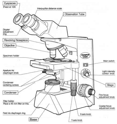
Microscope Parts & Functions - AmScope Main Microscope Parts and Functions. Head: The upper part of the microscope houses the eyepiece and objective lenses. Tube: Where the eyepieces are dropped in. Also, it connects the eyepieces to the objective lenses. Stage: The flat platform that supports the slides. Stage clips hold the slides in place.

Labels of a microscope and functions
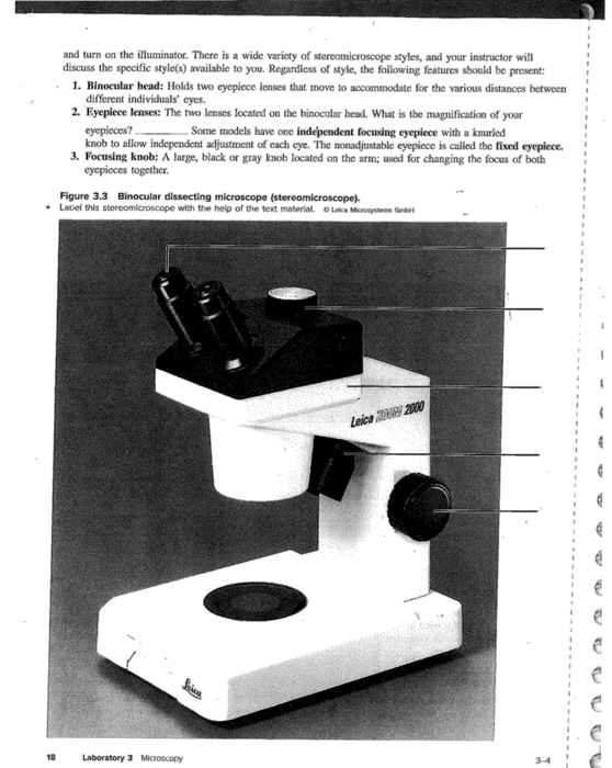
Microscope With Labeled Parts and Functions - 24 Hours Of Biology Optical parts and the functions The optical parts of the microscope are used to view, enlarge, and produce an image from a sample placed on a slide. These parts include Eyepiece: Eyepiece also contains ocular lens. It enhance the image of the viewer. This part is used for checking through the microscope. Eyepiece is found at the upper part of it. Parts of Stereo Microscope (Dissecting microscope) – labeled ... Unlike a compound microscope that offers a flat image, stereo microscopes give the viewer a 3-dimensional image that you can see the texture of a larger specimen. [In this image] Examples of Stereo & Dissecting microscopes. Major microscope brands (Zeiss, Olympus, Nikon, Amscope, Omano, Leica …) all produce stereomicroscopes. Parts of the Microscope with Labeling (also Free Printouts) Parts of the Microscope with Labeling (also Free Printouts) By Editorial Team March 7, 2022 A microscope is one of the invaluable tools in the laboratory setting. It is used to observe things that cannot be seen by the naked eye. Table of Contents 1. Eyepiece 2. Body tube/Head 3. Turret/Nose piece 4. Objective lenses 5. Knobs (fine and coarse) 6.
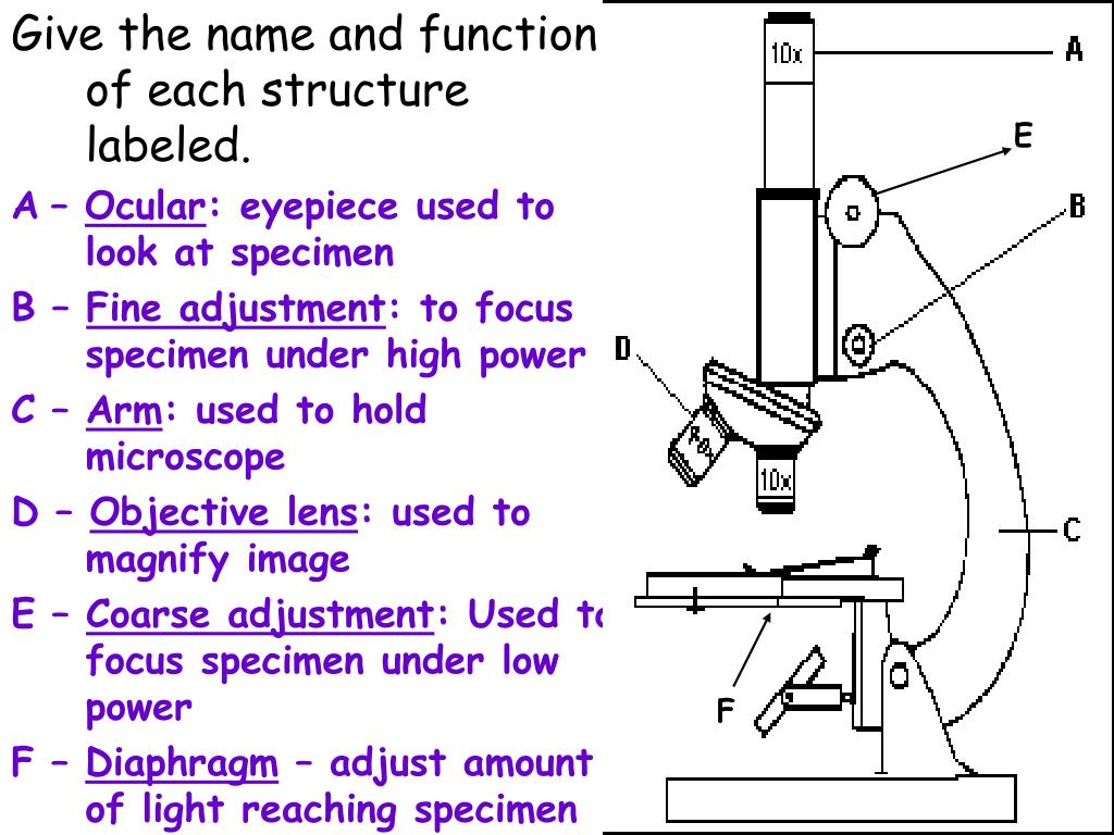
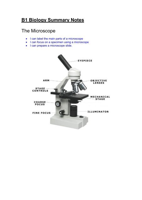
Labels of a microscope and functions. Compound Microscope Parts - Labeled Diagram and their Functions The eyepiece (or ocular lens) is the lens part at the top of a microscope that the viewer looks through. The standard eyepiece has a magnification of 10x. You may exchange with an optional eyepiece ranging from 5x - 30x. [In this figure] The structure inside an eyepiece. The current design of the eyepiece is no longer a single convex lens. Parts of the Microscope Label and Definition Diagram | Quizlet Start studying Parts of the Microscope Label and Definition. Learn vocabulary, terms, and more with flashcards, games, and other study tools. Home. Subjects. Explanations. ... Microscope Parts and Functions. 14 terms. soccer20022002. biology microscope parts. 12 terms. molini007. Sets found in the same folder. testes labeling. 8 terms ... Parts of a Microscope Labeling Activity - Storyboard That Create a poster that labels the parts of a microscope and includes descriptions of what each part does. Click "Start Assignment". Use a landscape poster layout (large or small). Search for a diagram of a microscope. Using arrows and textables label each part of the microscope and describe its function. Label Microscope Diagram - EnchantedLearning.com Using the terms listed below, label the microscope diagram. arm - this attaches the eyepiece and body tube to the base. base - this supports the microscope. body tube - the tube that supports the eyepiece. coarse focus adjustment - a knob that makes large adjustments to the focus. diaphragm - an adjustable opening under the stage, allowing ...
5 Types of Microscopes with Definitions, Principle, Uses, Labeled Diagrams It is used to visualize the living cells by creating a difference in contrast between the cells and water. It converts slight differences in refractive index and cell density into easily detectable variations in light intensity. It is useful for studying: Microbial motility Determining the shape of living cells Label the microscope — Science Learning Hub Jun 08, 2018 · All microscopes share features in common. In this interactive, you can label the different parts of a microscope. Use this with the Microscope parts activity to help students identify and label the main parts of a microscope and then describe their functions. Drag and drop the text labels onto the microscope diagram. If you want to redo an ... Parts of a microscope with functions and labeled diagram - Microbe Notes Microscopes are instruments that are used in science laboratories to visualize very minute objects such as cells, and microorganisms, giving a contrasting image that is magnified. Microscopes are made up of lenses for magnification, each with its own magnification powers. Compound Microscope Parts, Functions, and Labeled Diagram Compound Microscope Definitions for Labels. Eyepiece (ocular lens) with or without Pointer: The part that is looked through at the top of the compound microscope. Eyepieces typically have a magnification between 5x & 30x. Monocular or Binocular Head: Structural support that holds & connects the eyepieces to the objective lenses.
Microscope Types (with labeled diagrams) and Functions Microscopes aid in identifying viruses, bacteria and other microbial organisms and help the researchers in studying about diseases and find a cure to them. Researchers and pathologists examine several specimens a day to understand the underlying cause to a disease to enable doctors to give the right treatment to their patients Parts of a Compound Microscope and Their Functions - NotesHippo It controls the amount and intensity of light that enters the microscope. It can be either an iris diaphragm or a disc diaphragm. Condenser: It's a lense that's hidden beneath the stage. The size of the light beam is controlled by it. It collects and directs light from the mirror to the objective lens. Parts of a Microscope - The Comprehensive Guide Step 1: Fully open field and condenser diaphragms and focus on specimen using x10 objective. Step 2: Fully close field diaphragm and adjust the condenser and focus so edges are as sharp as possible. Step 3: Use screws at front of condenser to centre field diaphragm and open field diaphragm to fill view. Step 4: Remove eyepiece and close down ... Simple Microscope - Parts, Functions, Diagram and Labelling Mirror - A simple microscope has a plano-convex mirror and its primary function is to focus the surrounding light on the object being examined. Lens - The biconvex lens is placed above the stage and its function is to magnify the size of the object being examined.
Microscope Diagram to Print
Microscope parts — Science Learning Hub In this activity, students identify and label the main parts of a microscope and describe their function. By the end of this activity, students should be able to: identify the main parts of a microscope. describe the function of the different parts of a microscope. Download the Word file (see link below) for: background information for teachers.

how to use Microscope,describing different Parts - YouTube
ch 8 mastering biology Flashcards | Quizlet Looking through a light microscope at a dividing cell, you see two separate groups of chromosomes on opposite ends of the cell. New nuclear envelopes are taking shape around each group. The chromosomes then begin to disappear as they unwind.
PPT - Microscope Review PowerPoint Presentation, free download - ID:3102854
LAS X Industry Microscope software for Industry | Products ... Measure parameters, such as the length, area, diameter, angle, or perimeter of objects you mark with adjustable tracing lines, drawing directly in the live images. Add labels for easy analysis. Apply measurements to several images to determine statistical trend and compare data in measurement templates.

Compound Light Microscope
Electron microscope - Wikipedia An electron microscope is a microscope that uses a beam of accelerated electrons as a source of illumination. As the wavelength of an electron can be up to 100,000 times shorter than that of visible light photons , electron microscopes have a higher resolving power than light microscopes and can reveal the structure of smaller objects.
Microscope Drawing And Label at GetDrawings | Free download
Microscope, Microscope Parts, Labeled Diagram, and Functions Microscopes magnify or enlarge small objects such as cells, microbes, bacteria, viruses, microorganisms etc. at a viewable scale for examination and analysis. Microscopes consist of one or more magnification lenses to enlarge the image of the microscopic objects placed in the focal plane.

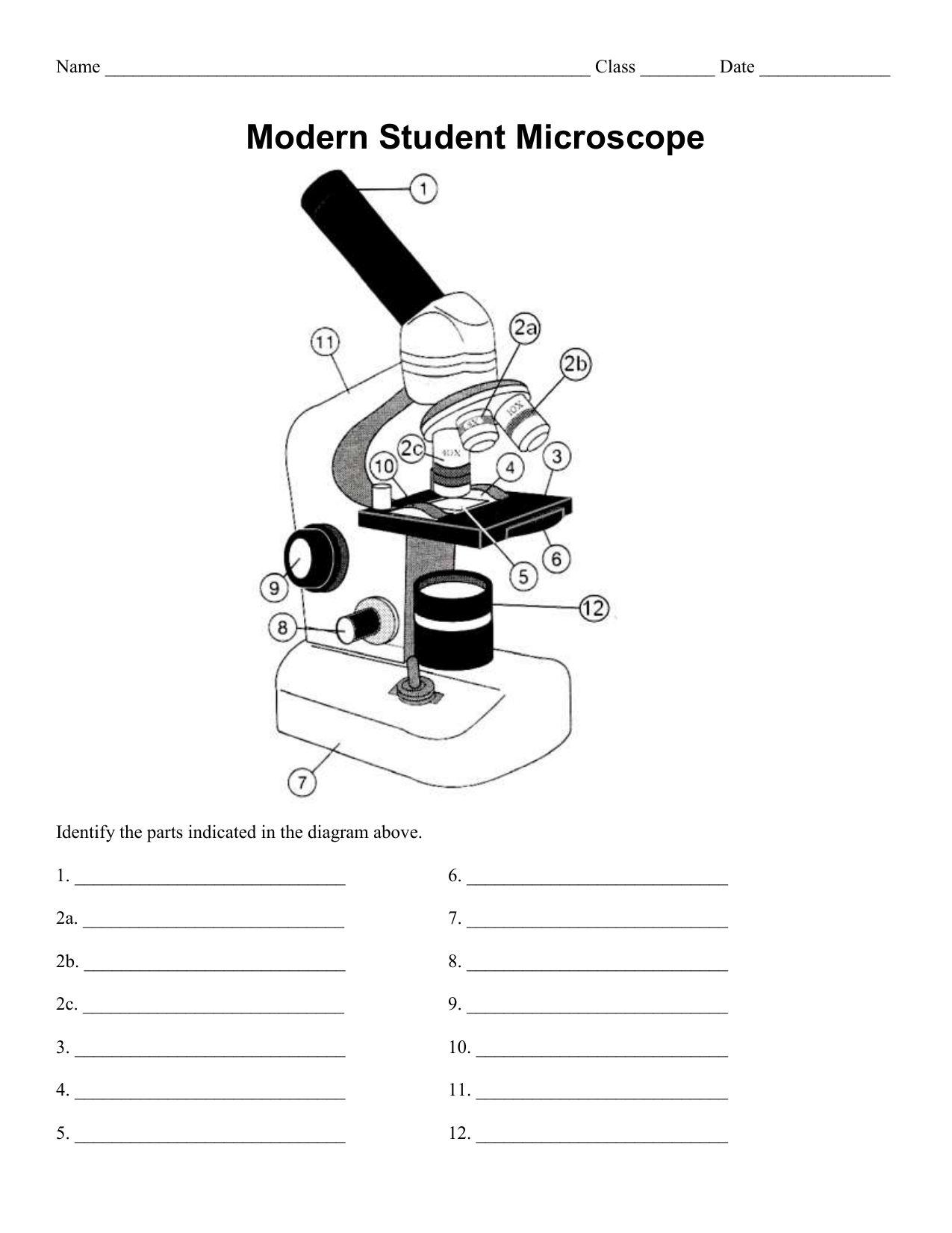
31 Label The Indicated Parts Of The Microscope - Labels Database 2020
Microscope Parts and Functions First, the purpose of a microscope is to magnify a small object or to magnify the fine details of a larger object in order to examine minute specimens that cannot be seen by the naked eye. Here are the important compound microscope parts... Eyepiece: The lens the viewer looks through to see the specimen.
32 Label Parts Of A Microscope - Labels For You
Compound Microscope Parts, Function, & Diagram - Study.com The base of the compound light microscope is the bottom portion of the compound microscope. It functions to support the entire compound microscope. The base can be set on a table or lab bench, and ...

Label the diagram of the microscope and explain the role of each part: - Brainly.in
22 Parts Of a Microscope With Their Function And Labeled Diagram A light microscope is a type of microscope that commonly uses visible light and a system of lenses to generate magnified images of small objects whereas electron microscope is a microscope that uses a beam of accelerated electrons as a source of illumination. It is a special type of microscope with a high resolution of images.

animal cell diagram (graphic) with organelles labelled | Animal cell, Cell biology, Animal cell ...
Microscope labeling and functions Flashcards | Quizlet Start studying Microscope labeling and functions. Learn vocabulary, terms, and more with flashcards, games, and other study tools.
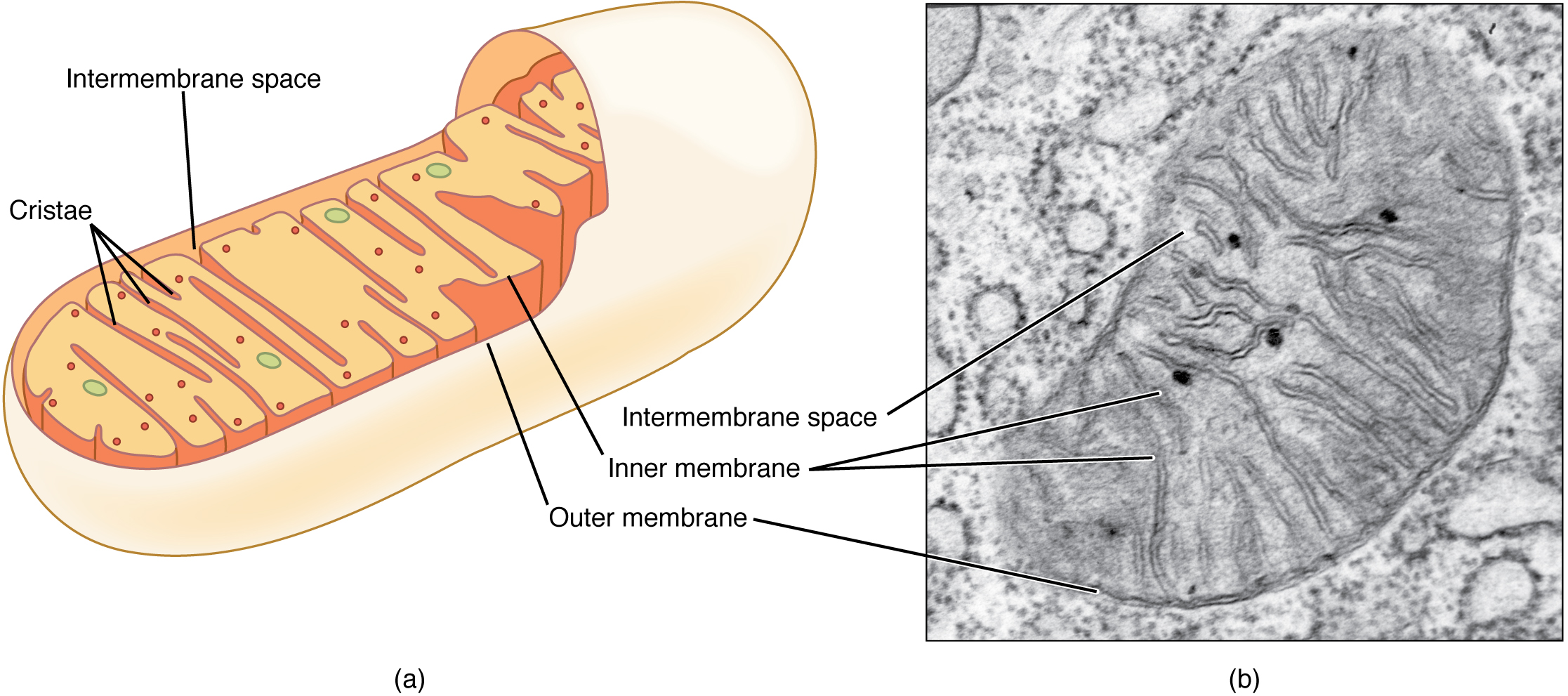
This figure shows the structure of a mitochondrion. The inner and outer membrane, the cristae ...
PDF Label parts of the Microscope Label parts of the Microscope: . Created Date: 20150715115425Z
Microscope labeling, modern and classical types
Microscope Parts, Function, & Labeled Diagram - slidingmotion Microscope parts labeled diagram gives us all the information about its parts and their position in the microscope. Microscope Parts Labeled Diagram The principle of the Microscope gives you an exact reason to use it. It works on the 3 principles. Magnification Resolving Power Numerical Aperture. Parts of Microscope Head Base Arm Eyepiece Lens

31 Label The Indicated Parts Of The Microscope - Labels For Your Ideas
List: Parts of a Microscope and their Function | Pathwooded It's the part of your microscope that you will look through to study objects and specimens. The eyepiece or ocular lens of a light microscope usually has a magnification level of 10x or 15x, but this can vary depending upon the microscope that you buy. Some microscope eyepieces have an adjustable magnification level. Microscope Tube
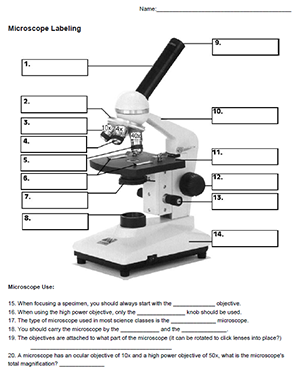
Microscope Labeling
Confocal Microscopy - an overview | ScienceDirect Topics John M. Murray, in Methods in Cell Biology, 2013 Abstract. Confocal microscopes are in principle well suited for quantitative imaging. The 3D fluorophore distribution in a specimen is transformed by the microscope optics and detector into the 2D intensity distribution of a digital image by a linear operation, a convolution.

Simple Microscope Labelled Diagram - Micropedia
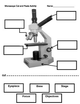
A Study of the Microscope and its Functions With a Labeled Diagram ... Compound Microscope Parts and Functions Body Tube - It is the part of the microscope that holds the eyepiece. Arm - The arm connects the body tube to the base. The user must hold this part in order to move the microscope from one place to another.
Microscope Labeling Activity by Interactive Creations | TpT
Introduction to three-dimensional image processing skimage.exposure contains a number of functions for adjusting image contrast. These functions operate on pixel values. Generally, image dimensionality or pixel spacing does not need to be considered. Gamma correction, also known as Power Law Transform, brightens or darkens an image. The function \(O = I^\gamma\) is applied to each pixel in the ...
31 Label The Indicated Parts Of The Microscope - Labels Database 2020
Label The Parts Of A Microscope Teaching Resources | TpT Hashtag Teached. 4.9. (10) $3.00. $2.00. PDF. Check out this well-organized Microscope label and describe worksheet. Part I is a visual that students will label and it corresponds with Part II where they will describe the function of those parts. This is a great worksheet that can easily be used for classwork, group work, homework, assessments ...

8 Best Images of Lens Diagram Worksheet - Microscope with Labeled Parts, Label Eye Parts ...
Parts of the Microscope with Labeling (also Free Printouts) Parts of the Microscope with Labeling (also Free Printouts) By Editorial Team March 7, 2022 A microscope is one of the invaluable tools in the laboratory setting. It is used to observe things that cannot be seen by the naked eye. Table of Contents 1. Eyepiece 2. Body tube/Head 3. Turret/Nose piece 4. Objective lenses 5. Knobs (fine and coarse) 6.
Post a Comment for "43 labels of a microscope and functions"