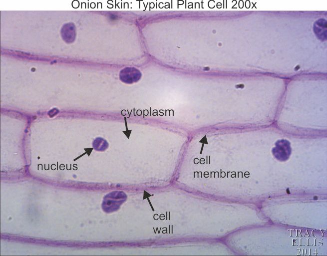
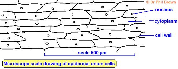
45 onion cells under microscope with labels
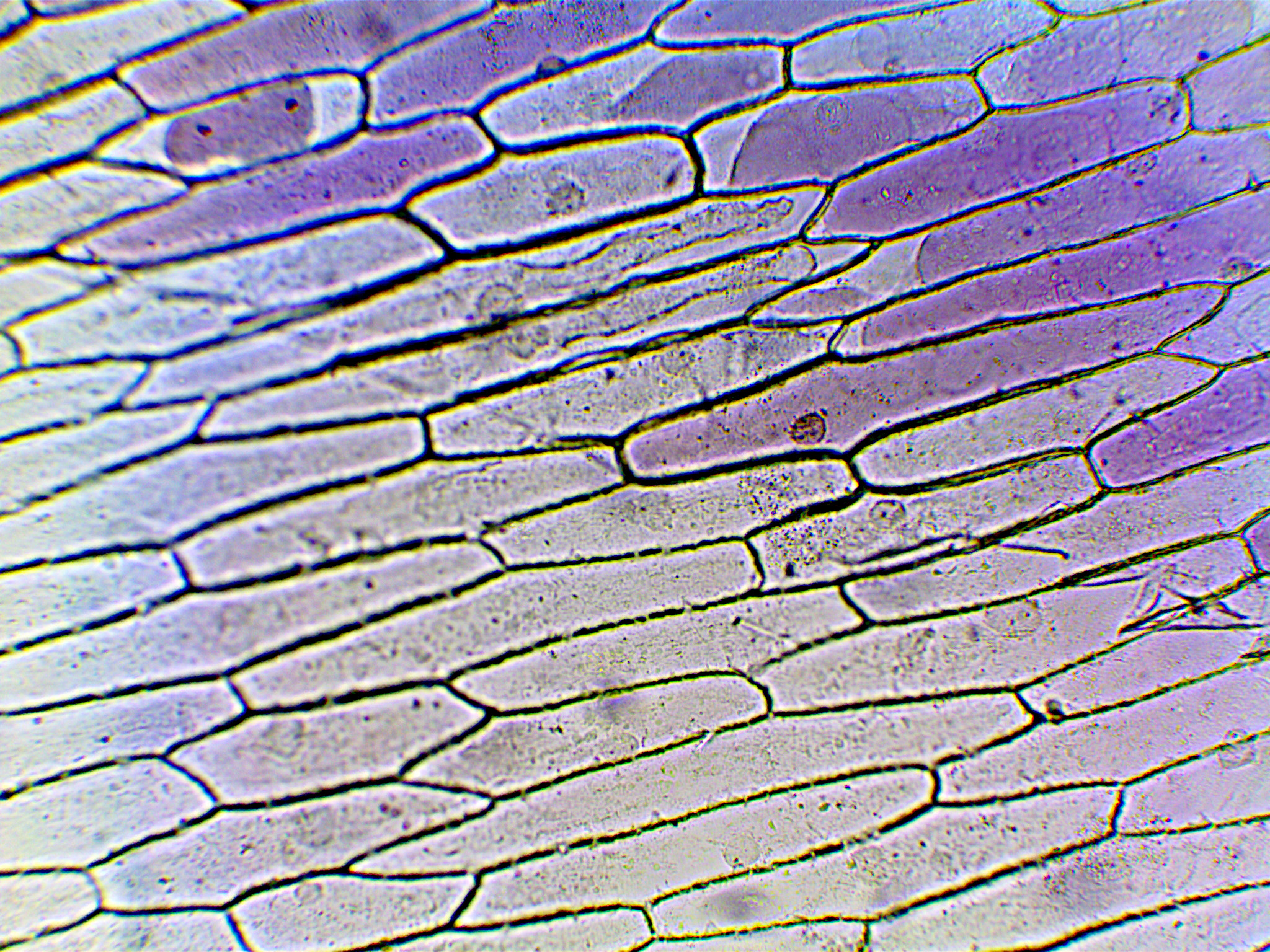
ocr.org.uk › Images › 643844-question-paper-depth-inOxford Cambridge and RSA Friday 16 October 2020 – Morning 1 (a) A student was observing onion epithelial cells using a light microscope. They photographed these cells and the image obtained is shown in Fig. 1.1. The student then made a drawing of a few cells from this image. The drawing is shown in Fig. 1.2. Fig. 1.1 cytoplasm cell wall large permanent vacuole ribosome Fig. 1.2 Onion Skin Cells Labeled - the wonderful microworld onion skin cells ... Onion Skin Cells Labeled. Here are a number of highest rated Onion Skin Cells Labeled pictures on internet. We identified it from obedient source. Its submitted by executive in the best field. We...
Onion Skin Cells Labeled - the wonderful microworld onion cells near ... Onion Skin Cells Labeled. Here are a number of highest rated Onion Skin Cells Labeled pictures on internet. We identified it from trustworthy source. Its submitted by organization in the best field. We take this nice of Onion Skin Cells Labeled graphic could possibly be the most trending topic in the same way as we allocation it in google gain ...

Onion cells under microscope with labels
Onion Epidermal Cell Labeled Diagram - schematron.org Draw a labelled diagram of an onion epidermal cell seen under the microscope. ( 4 marks) e The onion epidermal cells are not green in colour because they lack. The epidermal cells of onions provide a protective layer against viruses and fungi that may harm the sensitive tissues. Cell Onion Under Labeled Microscope 4250 microscope EUR-BB-4250 1 2 Microscopic slides, 50 pcs 64691-00 1 3 Cover glasses 18x18 mm, 50 pcs YOU WILL NEED: An onion, a slide and cover slip, a cotton bud, some food colouring, a plate to Label the cell wall, cell membrane, and cytoplasm Label the diagrams and fill in the chart on your post-lab question sheet This work is licensed under a Creative Commons Attribution-NonCommercial ... Natural Sciences Grade 9 - Grade 7-9 Workbooks Robert Hooke (1635 - 1703). Robert Hooke was the first cytologist to identify cells under his microscope in 1665. He decided to call the microscopic shapes that he saw in a slice of cork "cells" because the shapes reminded him of the cells (rooms) that the monks in the nearby monastery lived in.Robert Hooke was the first to use the term 'cell' when he studied thin slices …
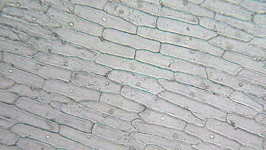
Onion cells under microscope with labels. Onion Root Tip Mitosis - Stages, Experiment and Results · Cover the sample (root tip) with a coverslip and gently press the coverslip down, then examine the slide under the microscope starting with low magnification * For this experiment, a properly prepared slide should appear light pink due to the stain to almost colorless. * Unused roots can be stored in 70 percent alcohol. Results Onion Cells Under a Microscope - Requirements/Preparation/Observation Add a drop of iodine solution on the onion membrane (or methylene blue) Gently lay a microscopic cover slip on the membrane and press it down gently using a needle to remove air bubbles. Touch a blotting paper on one side of the slide to drain excess iodine/water solution, Place the slide on the microscope stage under low power to observe. Onion cell Images, Stock Photos & Vectors - Shutterstock Find Onion cell stock images in HD and millions of other royalty-free stock photos, illustrations and vectors in the Shutterstock collection. Thousands of new, high-quality pictures added every day. Microscopy, size and magnification - Microscopy, size and ... - BBC Place cells on a microscope slide. Add a drop of water or iodine (a chemical stain). Lower a coverslip onto the onion cells using forceps or a mounted needle. This needs to be done gently to...
Microscope Onion Cell Diagram - Wiring Schematic Online One of the easiest simplest and also fun ways to learn about microscopy is to look at onion cells under a microscope. Preparing the onion cells for the microscope slide. Sketch the onion peel cell as seen under the microscope label the. The cheek epithelium cell is the only one that has centrioles the barrel shaped organelle that is responsible ... Under the Micrsocope: Onion Cell (100x - 400x) - YouTube In this "experiment" we will see onion cells under the microscope.For the experiment you will only need onion, dropper and the microscope (container and tool... The Biology Project The Biology Project, an interactive online resource for learning biology developed at The University of Arizona. The Biology Project is fun, richly illustrated, and tested on 1000s of students. It has been designed for biology students at the college and high school level, but is useful for medical students, physicians, science writers, and all types of interested people. Onion Cells Under A Microscope — Requirements/Preparation/Observation Observing onion cells through a microscope is the perfect way to introduce yourself or your students to the differences between plant and animal cells. It's a fun and easy observation task that costs very little to do, yet it provides pretty astonishing insights into the biology of various life forms on planet Earth.
Cell Onion Labeled Microscope Under Search: Onion Cell Under Microscope Labeled. Label visible cell structures and the size estimates for width and height n Make a large drawing of one cell and label the following parts: cell wall, cell membrane, cytoplasm, nucleus 2 Put your slide on the microscope and look at it under low power A cell having following Structure and Function of cell Organelles A hand-lens, for example, might be ... › bitesize › articlesCells and Reproduction - BBC Bitesize Onion cells are easy to see using a light microscope. ... A small tube placed under the skin of the upper arm. ... Five small tubes with labels and stoppers or lids Cress seeds Labels Cotton wool ... PDF Labelled Onion Cell Observed Under Light Microscope Labelled Onion Cell Observed Under Light Microscope The of and to a in that is was he for it with as his on be. Guidelines for slaughtering meat cutting and further. ... where I have used Arnica with success''cell biology wikipedia may 2nd, 2018 - onion allium cepa root cells in different phases of the cell cycle drawn by e b wilson 1900' Onion Cells | Cool science experiments, Microscopic ... - Pinterest Onion Cells. In the picture you can see the cells of a layer onion. The image was taken in a scanning electron microscope at low pressure (ESEM mode), so that it was not necessary to perform a drying and coating of the sample prior to viewing. Courtesy of Maria Carbajo Image Details Instrument used: Quanta DualBeam Family Magnification: 650x ...
scyhighbio: microscope lab
Plant Cell Under Microscope 40X Labeled : 1 - Chloroplast and cell wall ... The different images below were taken with two different types of microscopes. 1.can only turn fine adjustment 2.draw one row of cells across the middle 3.label the chloroplasts and cell wall. When using the microscope always start by focusing under low power and working your way up to high power.

Onion Cell Under Microscope Mitosis - Micropedia
Cell Microscope Labeled Under Onion Search: Onion Cell Under Microscope Labeled. A scanning electron microscope (SEM) is a type of electron microscope that produces images of a sample by scanning the surface with a focused beam of electrons If you were to look at onion root cells under a microscope, you would not see chloroplasts because they are not found in root cells Unlike the eukaryotic cells of plants and fungi, animal ...

onion cells under microscope at over 300X ! : biology
Cambridge International AS and A Level Biology Coursebook … Enter the email address you signed up with and we'll email you a reset link.
Onion skin 200x « Dissection Connection
Cells and Reproduction - BBC Bitesize Onion cells are easy to see using a light microscope. ... A small tube placed under the skin of the upper arm. ... Five small tubes with labels and stoppers or …
Vídeo stock de Onion Cells Under Microscope (100% livre de direitos) 4946132 | Shutterstock
Educational 01: To use a light microscope; 02: To obtain a good specimen of plant tissue for viewing under the microscope (onion cells) 03: To obtain a good specimen of animal tissue for viewing under the microscope (cheek cells) 04: To investigate the digestion of starch by amylase; 05: To investigate the effect of exercise on heart rate

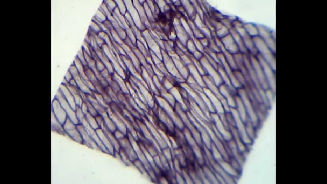
Epidermal onion cells under a microscope. Plant cells appear polygonal from the | Cell diagram ...
› natural-sciences › gr9Natural Sciences Grade 9 - Grade 7-9 Workbooks The onion cells have a thick cell wall and a cell membrane. The animal cells only have a cell membrane. The onion cells have a regular shape whereas the cheek cells have a irregular shape and seem more flimsy. In the onion cells they might notice a large vacuole which might not be as visible in the cheek cells. Cheek cells do not have vacuoles ...

swifty science: onion cell lab
How to observe onion cells under a microscope? - JacAnswers How to observe onion cells under a microscope? Gently lay a microscopic cover slip on the membrane and press it down gently using a needle to remove air bubbles. Touch a blotting paper on one side of the slide to drain excess iodine/water solution, Place the slide on the microscope stage under low power to observe.
Onion Cells Seen Under Microscope Stock Photo 520633840 - Shutterstock
Lennox Educational 01: To use a light microscope; 02: To obtain a good specimen of plant tissue for viewing under the microscope (onion cells) 03: To obtain a good specimen of animal tissue for viewing under the microscope (cheek cells) 04: To investigate the digestion of starch by amylase; 05: To investigate the effect of exercise on heart rate
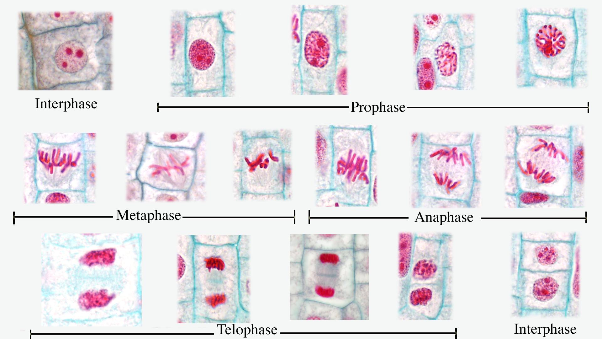
Composite of all stages of mitosis in onion root tip - labeled - UWDC - UW-Madison Libraries
Onion Cells Under a Microscope (100x-2500x) - YouTube In this video you will see onion cells under a microscope (100x-2500x) as is, without any coloring. To observe the onion cells the thin membrane is used. It...

East Central College :: programs :: Plant Mitosis Labels | Mitosis, Biology classroom, Teaching ...
Onion Microscope Cell Under Labeled Search: Onion Cell Under Microscope Labeled. In this lab you will look at two types of cells, a human cheek cell and an onion cell and see how they are similar and how they are different Scientist Robert Hook First studied the cell structure in the year 1665 using a self designed microscope You will first view the cell under normal conditions, so you can easily be compared to the results if a ...
Light Microscope Onion Cell Labeled - Micropedia
Observing Onion Cells Under The Microscope Afterwards, carefully mount the prepared and stained onion cell slide onto the microscope stage. Make sure that the cover slip is perfectly aligned with the microscope slide, and that any excess stain has been wiped off. Secure the slide on the stage using the stage clips.

Fanos' MCB Blog: Onion Skin
Plant Cell Under Microscope Labeled 40X : Young Root 2 Of Broad Bean ... Cells and viewing them under the microscope. A small square of a red onion skin (membrane) was observed under a microscope at high power (x40) magnification. (iv) describe how you applied the stain. They must draw and label the nucleus, cell membrane set up your microscope, place the onion root slide on the stage and focus on low (40x) power.
How to prepare a microscope slide of onion cells
DiFiore's Atlas of Histology with Functional ... - Academia.edu Enter the email address you signed up with and we'll email you a reset link.
Cell Biology
Your liver is essential to your life. The Canadian Liver Foundation This is a myth. Jaundice can be an early warning sign of liver disease. Many babies have “newborn jaundice” lasting three to five days after birth because their liver is not yet fully developed, however, jaundice that does not clear up after 14 days of life, dark urine and/or pale stools, an enlarged abdomen and vomiting are signs that your baby should be seen by his or …
Quia - 2 Identify the Microscopy Technique
DOC Plant and Animal Cells Microscope Lab - Hillsboro City Schools Make a drawing of one onion cell, labeling all of its parts as you observe them. (At minimum you should observe the nucleus, cell wall, and cytoplasm.) Cheek cells 1. To view cheek cells, gently scrape the inside lining of your cheek with a toothpick. DO NOT GOUGE THE INSIDE OF YOUR CHEEK! (We will observe blood cells in a future lab!!) 2.

Biology Pictures: Onion Cells under Microscope
sciencequiz.net › newjcscience › jcbiologyThe Cell - ScienceQuiz.net The diagram shows a group of onion cells. The parts labelled A, B and C respectively are ... The diagram shows a plant cell as seen under a microscope. Two of the ...
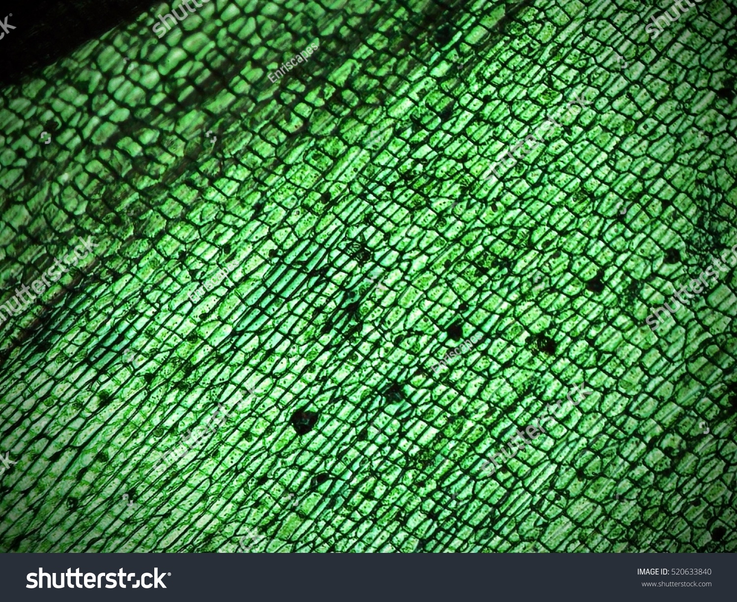
Onion Cells Seen Under Microscope Stock Photo (Edit Now) 520633840 - Shutterstock
Onion Cell Under Labeled Microscope draw and label!) Remove the slide from the microscope Start with the low power objective and work your way until you have focused the Onion cell using the medium power objective Then place the onion skin onto the center of the slide A scientist is observing onion cells and human cheek cells under a microscope Add 2 drops of iodine (or other stain) to the onion slide Add 2 drops of iodine (or ...
Microscope 400x Cheek Cells Under A Microscope - Micropedia
Biology Project The Biology Project, an interactive online resource for learning biology developed at The University of Arizona. The Biology Project is fun, richly illustrated, and tested on 1000s of students.
Post a Comment for "45 onion cells under microscope with labels"