41 ribosome diagram with labels
DNA Labeling: Transciption and Translation - The Biology Corner This worksheet shows a diagram of transcription and translation and asks students to label it; also includes questions about the processes. Name: _____ ... How does the ribosome know the sequence of amino acids to build? 12. What is the difference between a codon and an anticodon? Animal Cell Diagram | Science Trends The diagram, like the one above, will include labels of the major parts of an animal cell including the cell membrane, nucleus, ribosomes, mitochondria, vesicles, and cytosol. The cells of animals are the basic structural units for the wide variety of life we see in the animal kingdom.
Active Ribosome Profiling with RiboLace - PubMed Ribosome profiling, or Ribo-seq, is based on large-scale sequencing of RNA fragments protected from nuclease digestion by ribosomes. Thanks to its unique ability to provide positional information about ribosomes flowing along transcripts, this method can be used to shed light on mechanistic aspects … Active Ribosome Profiling with RiboLace

Ribosome diagram with labels
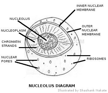
Campbell Ap Biology Mastering Biology Chapter 17 Course Work Drag the white and purple labels to the white targets to indicate what each mutant mRNA codon codes for. (You will probably need to consult the codon table for mRNA .) Drag the pink labels to the pink targets to indicate the type of mutation. Drag the blue labels to the blue targets to indicate the effect on the polypeptide's primary structure. Structure of Ribosome - Biology Wise Diameter of Ribosome is 20nm. Their composition can be divided into two parts - 2/3 part of r-RNA (ribosomal RNA) and 1/3 part RNP (Ribosomal protein or Ribonuclep protein). Polypeptide chain is fabricated by translating mRNA (messenger RNA) with the aid amino acids that tRNA (transfer RNA) delivers. Nuclear envelope - Wikipedia The nuclear envelope is punctured by around a thousand nuclear pore complexes, about 100 nm across, with an inner channel about 40 nm wide. The complexes contain a number of nucleoporins, proteins that link the inner and outer nuclear membranes.
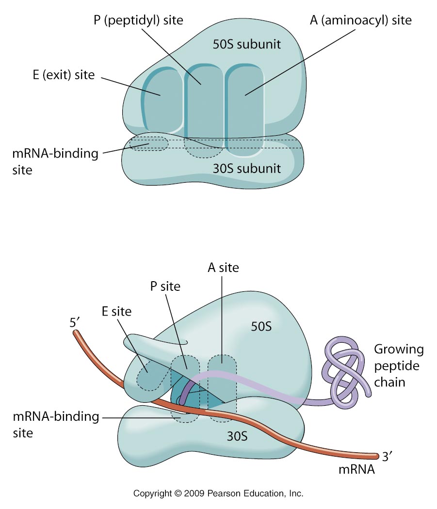
Ribosome diagram with labels. Oxford Cambridge and RSA Friday 16 October 2020 – Morning The teacher stated that two of the labels on the drawing Fig. 1.2 were incorrect, and also that it was a poor quality biological drawing. (i) Identify one incorrect label and explain your answer. Incorrect label ..... Ribosome - Wikipedia The ribosomal proteins and rRNAs are arranged into two distinct ribosomal pieces of different sizes, known generally as the large and small subunit of the ribosome. Ribosomes consist of two subunits that fit together (Figure 2) and work as one to translate the mRNA into a polypeptide chain during protein synthesis (Figure 1). Ribosome Images - Center for Molecular Biology of RNA Ribosome Images. All fullsize images are 300 pixels/inch and suitable for high resolution reproduction. All TIFF images use the CMYK color gamut, are are LZW compressed. All JPEG images use the RGB color gamut and have minimal compression. Structure of Ribosome (With Diagram) - Biology Discussion A bacterial ribosome is about 250 nm in diameter and consists of two subunits, one large and one small. Both subunits consist of one or more molecules of rRNA and an array of ribosomal proteins. ADVERTISEMENTS: Association of two subunits is called mono-some. The structure of prokaryotic ribosome is given in the figure 8.2 B.
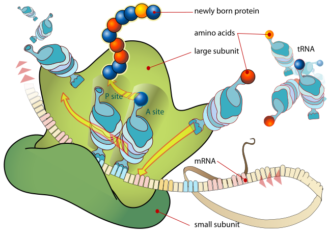
Ribosome and protein synthesis, diagram - Science Photo Library Ribosome and protein synthesis, diagram. C029/3019. Rights Managed. 50.0 MB (867.0 KB compressed) 4827 x 3620 pixels. 40.9 x 30.7 cm ⏐ 16.1 x 12.1 in (300dpi) This image is not available for purchase in your country. Please contact your Account Manager if you have any query. Request Price Add To Basket. Solved In the following diagram of a ribosome, assign the | Chegg.com in the following diagram of a ribosome, assign the correct labels. 5' end of the mrna growing polypeptide a trna attached to a single amino acid ontors here large subunit atrna attached to a polypeptide is found in this area of the nibosome a trna that is not attached to anything exits hore 3' end of the mrna a trna moleculo mossenger rna being … Biuret test - Wikipedia The biuret (IPA: / ˌ b aɪ j ə ˈ r ɛ t /, / ˈ b aɪ j ə ˌ r ɛ t /) test, also known as Piotrowski's test, is a chemical test used for detecting the presence of peptide bonds.In the presence of peptides, a copper(II) ion forms mauve-colored coordination complexes in an alkaline solution. protein synthesis diagram labeled - TheFitnessManual Switch RNAs (tRNAs) deliver amino acids to the ribosome. - "protein synthesis diagram labeled" tRNAs are additionally RNA polymers. They're typically between 75 and 90 RNA nucleotides lengthy. However in contrast to mRNAs, that are linear, hydrogen bonding between nucleotides inside a tRNA causes it to fold up.
Science S1 Flashcards | Quizlet The strand of RNA moves to the ribosome. The DNA double helix unzips. ... The diagram shows a chicken embryo and a human embryo. ... Ribosome and protein synthesis, diagram - Science Photo Library Diagram showing protein synthesis in cells (translation). Messenger ribonucleic acid (mRNA, blue with coloured nucleotides) is read by a ribosome (pink). The molecules of transfer RNA (tRNA, key-shaped) each bring an amino acid (orange dot) to bind to the ribosome's protein synthesis site. Bacteria in Microbiology - shapes, structure and diagram Bacterial spores. Bacterial endospores layers. Bacteria cells are the smallest living cells that are known; even though viruses are smaller than bacteria, viruses are not living cells. There are different types of bacteria with various sizes, shapes, and structures. The bacteria shapes, structure, and labeled diagrams are discussed below. Ribosomes: Structure, Composition, and Assembly (With Diagram) Ribosomes in the cytoplasm of eukaryotic cells have a sedimentation coefficient of about 80 S (MW about 4.5 x 10 6) and are composed of 40 S and 60 S subunits. In prokaryotic cells, ribosomes are typically about 70 S (MW about 2.7 x 10 6) and are formed from 30 S and 50 S subunits.

Ribosome - Definition, Function and Structure | Biology Dictionary
Animal Cells: Labelled Diagram, Definitions, and Structure Ribosomes Ribosomes create proteins. They can float freely in the cytoplasm or can be attached to the nuclear envelope. They create proteins by assembling amino acids into polypeptides. As the ribosomes build an amino acid chain, the chain is pushed into the endoplasmic reticulum.

Understanding the Relationship Between Ribosome Structure and Function ...
Solved The ribosome in the diagram is in the process of | Chegg.com The ribosome in the diagram is in the process of synthesizing a protein using directions transcribed from the DNA. Use the labels to identify each of the structures involved in translation and protein synthesis. Question: The ribosome in the diagram is in the process of synthesizing a protein using directions transcribed from the DNA.

Organelles, Enzymes and Other Interesting Small Things: A Guide to AS ...
Labeled Plant Cell With Diagrams | Science Trends The ribosomes are created in the nucleolus of the cell. Ribosomes are made out of two smaller subunits, a large ribosomes subunit and a small ribosomal subunits. The transfer RNA or tRNA encodes the correct series of genetic instructions into the mRNA or messenger RNA, which is what ensures that the right proteins are created.
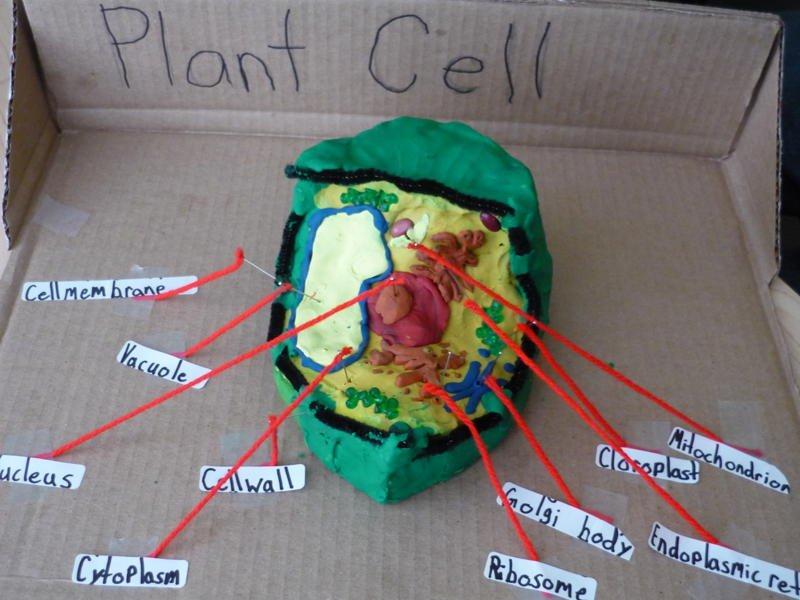
3d Plant Cell with labels : Biological Science Picture Directory ...
Mastering Quiz: Chapter 7A Microbial Genetics Flashcards | Quizlet c. mRNA binds to a ribosome in the cytoplasm. d. A molecule of RNA is formed based on the sequence of nucleotides in DNA. ... Drag the correct labels under the diagrams to identify the events of RNA processing. Drag the labels onto the diagram to identify how nucleotides pair up. Labels can be used once, more than once, or not at all.

71 best Cell Cycle images on Pinterest
Ribosome - Definition, Function and Structure | Biology Dictionary A. Ribosomes translate the 4 base language of DNA into the 20 base language of proteins, allowing for many more combinations. B. The 4 different nucleobases of DNA can be recombined endlessly to produce new proteins. C. Ribosomes can modify proteins with carbohydrates to make them unique. Answer to Question #2 3.

Ribosome - Cell (Biology) M.4
Ribosomes Images Stock Photos, Pictures & Royalty-Free Images - iStock Ribosomes vector illustration. Anatomical and medical labeled scheme with tRNA, Amino acid, protein, cell, small and large subunit, mRNA. Explained closeup diagram. ribosomes images stock illustrations
Ribosome Diagram - ClipArt Best
A Labelled Diagram Of Mitochondria with Detailed Explanation It is a viscous or a gel-like fluid containing a mixture of enzymes, ribosomes, inorganic ions, mitochondrial DNA, nucleotide cofactors, and organic molecules. It is involved in the cellular respiration and production of ATP molecules. Cristae The inner layer, surrounded by the folds of the mitochondrial matrix are collectively referred to Cristae.
The Cell
What Are Ribosomes? - Definition, Structure and its Functions Ribosomes are located inside the cytosol found in the plant cell and animal cells. The ribosome structure includes the following: It is located in two areas of cytoplasm. Scattered in the cytoplasm. Prokaryotes have 70S ribosomes while eukaryotes have 80S ribosomes. Around 62% of ribosomes are comprised of RNA, while the rest is proteins.
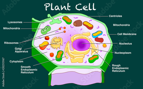
Plant cell structure. Annotated parts diagram. Anatomy with organelles ...
Prokaryotic Cells - BioNinja Ribosomes – complexes of RNA and protein that are responsible for polypeptide synthesis (prokaryote ribosome = 70S) Cell membrane – Semi-permeable and selective barrier surrounding the cell Cell wall – rigid outer covering made of peptidoglycan; maintains shape and prevents bursting (lysis)

Ribosome - Wikipedia
Ribosomes vector illustration - VectorMine Most Vector Editing Software. 3. High-resolution JPG image. 3800 x 3965 px. License terms in short: Use for everything except reselling item itself. Read a full license here. Description: Ribosomes vector illustration. Anatomical and medical labeled scheme with tRNA, Amino acid, protein, cell, small and large subunit, mRNA.
3.5/7.4 Translation | i am so
SA EXAM PAPERS The correct labels for parts X, Y and ... 2.1.1 Identify the stage of protein synthesis that is shown in the diagram above. (1) ... Protein synthesis at the ribosome .

Protein Synthesis
Ribosomes- Definition, Structure, Functions and Diagram Ribosomes Definition The ribosome word is derived - 'ribo' from ribonucleic acid and 'somes' from the Greek word 'soma' which means 'body'. Ribosomes are tiny spheroidal dense particles (of 150 to 200 A0 diameters) that are primarily found in most prokaryotic and eukaryotic. They are sites of protein synthesis.
The Control Center - Cells & Organelles
Ribosomes Vector Illustration. Anatomical and Medical Labeled Scheme ... Ribosomes vector illustration. Anatomical and medical labeled scheme. Explained closeup diagram.. Illustration about amino, educational, biogenesis, labeled, golgi, body - 122097933
Post a Comment for "41 ribosome diagram with labels"